Anatomy Of Ribs Posterior - Learn Muscle Anatomy: Serratus Posterior Superior and Inferior / It acts to depress the ribs.. Anteriorly, most are attached directly to the sternum. Each pair is numbered based on their attachment to the sternum, a bony process at the front of the rib cage which serves as an anchor point. Its function is to elevate the ribs. This ligament lies on the posterior of the body, while the sternocostal ligaments comprise the anterior aspect of the body. The typical ribs have a generalized structure, while the atypical ribs have variations on this structure.the typical ribs consist of two ends, a posterior or vertebral end, an anterior or sternal end, and an intervening portion identified as the body or shaft.
Anteriorly, most are attached directly to the sternum. Anatomy of ribs posterior : Home > human being > anatomy > skeleton > posterior view. The head of the rib is the end part closest to the vertebra with which it articulates. These muscle fibres extend in a posteroinferior direction and again pass in an oblique manner.
Easy Notes On 【Ribs】Learn in Just 4 Minutes! - Earth's Lab from www.earthslab.com Ribs anatomy, ligaments and clinical notes these pictures of this page are about:posterior rib anatomy. There are twelve pairs of ribs. The tubercle is a bony prominence located on the posterior side of each typical rib at the junction between the neck and the body. Common characteristics of the ribs (figs. It branches from the ileocolic artery and may branch further to the appendicular artery. Anatomy of ribs posterior : The lungs are a pair of cone‐shaped bodies that occupy the thorax. The nomenclature of the costal veins is the same as the arteries.
The ribs are elastic arches of bone, which form a large part of the thoracic skeleton.
In this video, you will learn the bony features of typical and atypical ribs. Gross anatomy there are 12 pairs of ribs which are separated by intercostal spaces. They articulate at the costochondral joints with some exceptions. Posterior rib anatomy anatomy drawing diagram from d1yboe6750e2cu.cloudfront.net the posterior abdominal wall is a musculoskeletal structure formed by the posterior abdominal muscles posteriorly by the lumbar vertebrae, muscles, and fascia. The tubercle is a bony prominence located on the posterior side of each typical rib at the junction between the neck and the body. There are twelve pairs of ribs. The ribs are attached to corresponding thoracic vertebrae posteriorly. Rib cage anatomy function britannica / in the anatomical position, the scapula overlies the second to seventh ribs on the posterolateral aspect of the chest wall. Rib cage, in vertebrate anatomy, basketlike skeletal structure that forms the chest, or thorax, and is made up of the ribs and their corresponding attachments to the sternum (breastbone) and the vertebral column.the rib cage surrounds the lungs and the heart, serving as an important means of bony protection for these vital organs.in total, the rib cage consists of the 12 thoracic vertebrae and. It branches from the ileocolic artery and may branch further to the appendicular artery. The eleven pairs of internal intercostal muscles are found posterior to the external intercostals. The lungs are a pair of cone‐shaped bodies that occupy the thorax. These pass from the inferior edge of the costal groove to the superior margins of the ribs below.
Each rib has two extremities, a posterior or vertebral, and an anterior or sternal, and an intervening portion—the body or shaft. The rib cage is a bony structure found in the chest (thoracic cavity). The sternocostal ligament is a broad, membranous band that originates from the anterior and posterior aspect of the sternal cartilage of the upper ribs and inserts to the posterior surfaces of the sternum. The flexible (hyaline) cartilage, makes the breathing process easier. These muscle fibres extend in a posteroinferior direction and again pass in an oblique manner.
Multiple Rib Fractures | Rib fracture from i.pinimg.com The ribs partially enclose and protect the chest cavity, where many vital organs (including the heart and the lungs) are located. The number is the same in both males and females. These muscle fibres extend in a posteroinferior direction and again pass in an oblique manner. Home > human being > anatomy > skeleton > posterior view. Anteriorly, most are attached directly to the sternum. Anatomy bones learning bone anatomy ask a biologist. The rib cage is a bony structure found in the chest (thoracic cavity). Lateral view of a pair of ribs articulating with the thoracic vertebrae.
Posterior rib anatomy anatomy drawing diagram from d1yboe6750e2cu.cloudfront.net the posterior abdominal wall is a musculoskeletal structure formed by the posterior abdominal muscles posteriorly by the lumbar vertebrae, muscles, and fascia.
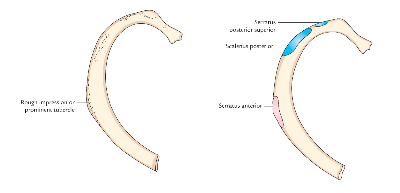
In contrast, in the cranial rib pair (s), the posterior rib (arrowhead) is higher than the anterior rib. Lateral view of a pair of ribs articulating with the thoracic vertebrae. The rib cage is a bony structure found in the chest (thoracic cavity). Home > human being > anatomy > skeleton > posterior view. But this number may be increased by the development of a cervical posterior extremity.—the posterior or vertebral extremity presents for examination a head. Serratus posterior superior and serratus posterior inferior. The anterior border of the lung is formed by the convergence of the mediastinal. The anatomy of the human ribs is made up of 24 ribs which are parted in 12 pairs (each on the left and right side of the chest wall), with the sternum, metasternum (the xiphoid process), and the costal cartilages all situated at the anterior of the chest wall, followed by the thoracic vertebrae on the posterior of the chest wall. The thoracic spine, composed of 12 segments, is the longest subsection of the vertebral column. Posterior rib anatomy anatomy drawing diagram from d1yboe6750e2cu.cloudfront.net the posterior abdominal wall is a musculoskeletal structure formed by the posterior abdominal muscles posteriorly by the lumbar vertebrae, muscles, and fascia. Common characteristics of the ribs (figs. All ribs are attached posteriorly to the thoracic vertebrae. Anteriorly, most are attached directly to the sternum.
Anatomy of ribs posterior : The sternum connects to the ribs by thin bands of cartilage called the costal cartilage. Ninja nerds!join us in this video where we show the sternum and rib articulation anatomy through the use of a model. These pass from the inferior edge of the costal groove to the superior margins of the ribs below. Posterior view of vertebrae anatomy in this image, you will find cervical vertebrae, thoracic vertebrae, lumbar vertebrae, sacrum, coccyx, spinal column, scapula, ribs, pelvic bone in it.
Rib Bones Anatomy - Anatomy Drawing Diagram from www.meddean.luc.edu In the anatomical position, the scapula overlies the second to seventh ribs on the posterolateral aspect of the chest wall. Anatomy bones learning bone anatomy ask a biologist. The ribs partially enclose and protect the chest cavity, where many vital organs (including the heart and the lungs) are located. Ninja nerds!join us in this video where we show the sternum and rib articulation anatomy through the use of a model. There are twelve (12) pairs of ribs and all articulate posteriorly with the thoracic vertebrae. In the inferior pair of ribs (i), the posterior rib (arrow) is slightly lower than the anterior rib. The lungs are a pair of cone‐shaped bodies that occupy the thorax. The sternum connects to the ribs by thin bands of cartilage called the costal cartilage.
The ribs are attached to corresponding thoracic vertebrae posteriorly.
Posterior rib anatomy anatomy drawing diagram from d1yboe6750e2cu.cloudfront.net the posterior abdominal wall is a musculoskeletal structure formed by the posterior abdominal muscles posteriorly by the lumbar vertebrae, muscles, and fascia. Posteriorly, the heads of the ribs interdigitate with the vertebrae and are numbered according. The typical ribs have a generalized structure, while the atypical ribs have variations on this structure.the typical ribs consist of two ends, a posterior or vertebral end, an anterior or sternal end, and an intervening portion identified as the body or shaft. All ribs are attached posteriorly to the thoracic vertebrae. Each rib has two extremities, a posterior or vertebral, and an anterior or sternal, and an intervening portion—the body or shaft. Gross anatomy there are 12 pairs of ribs which are separated by intercostal spaces. The posterior abdominal wall is a musculoskeletal structure formed by the posterior abdominal muscles posteriorly by the lumbar vertebrae, muscles, and fascia. The sternum connects to the ribs by thin bands of cartilage called the costal cartilage. / the ribs are the skeletal protection for the lungs and the chest cavity. The lungs are a pair of cone‐shaped bodies that occupy the thorax. In the inferior pair of ribs (i), the posterior rib (arrow) is slightly lower than the anterior rib. The anatomy of the human ribs is made up of 24 ribs which are parted in 12 pairs (each on the left and right side of the chest wall), with the sternum, metasternum (the xiphoid process), and the costal cartilages all situated at the anterior of the chest wall, followed by the thoracic vertebrae on the posterior of the chest wall. The eleven pairs of internal intercostal muscles are found posterior to the external intercostals.
The sternocostal ligament is a broad, membranous band that originates from the anterior and posterior aspect of the sternal cartilage of the upper ribs and inserts to the posterior surfaces of the sternum anatomy of ribs. Test your knowledge about the ribs anatomy here in vertebrate anatomy, ribs (latin:


0 Comments:
Posting Komentar